25 April 2024
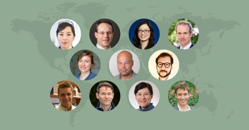
In the LEO Foundation’s latest round of Research Grants in open competition, 11 skin research projects are awarded a total of DKK 42 million. These projects range widely in their dermatological focuses, from the exploration of wound healing to the study of the herpes simplex virus, which infects the skin.
The LEO Foundation’s Research Grants program aims to support the best skin research projects worldwide which have the potential to better our understanding of the skin and its diseases. In its latest funding round, 11 skin research projects receive a total of DKK 42 million to generate novel and exciting dermatological insights.
The round attracted 39 reviewed applications requesting a total of DKK 132 million in funding.
A glance into the projects
New technology with the potential to revolutionize skin disease treatment
Among the 11 projects to receive a grant from the LEO Foundation is Associate Professor Niclas Roxhed’s, who hails from KTH Royal Institute of Technology in Sweden. Niclas Roxhed’s technology-focused project offers promise to potentially revolutionize the treatment of skin diseases, as he aims to look for new ways to deliver biopharmaceuticals to treat atopic dermatitis directly through the skin.
A great challenge for modern biological drug therapies is their inability to penetrate the skin’s outermost protective layers to deliver their large-molecule drugs to the deeper areas of the skin. The outermost layer of the skin is particularly tough and does not allow large molecules such as biological drugs to pass, therefore necessitating delivery methods like injections. Niclas Roxhed, however, takes on this challenge with his tailor-made ultra-sharp spiked microspheres that are able to transport the drugs and painlessly penetrate the skin.
With his funded project, Niclas Roxhed and his team will trial this technology in an atopic dermatitis model, with the results potentially forming the basis for highly effective topical treatment of skin diseases through creams, replacing the need for typical techniques such as injection.
A novel approach to tackling scleroderma
Assistant Professor Eliza Pei-Suen Tsou from the University of Michigan in the USA also receives a grant from the LEO Foundation, as she looks to pave the way for novel treatments for scleroderma – an autoimmune disease characterized by inflammation, scarring of tissues and organs, including the skin, and changes in blood vessels throughout the body.
More specifically, Eliza Pei-Suen Tsou places the spotlight on a process called senescence, as she hypothesizes that the aging process of dermal blood vessel endothelial cells might play a crucial role in scleroderma’s progression. Senescence can be described as a process by which a cell ages but fails to die, leading to a large buildup of these potentially dysfunctional senescent cells in the body. Such cells remain active, releasing harmful substances that can cause inflammation and damage in nearby healthy cells.
Having already determined that dermal endothelial cells from scleroderma patients function differently compared to healthy controls, Eliza Pei-Suen Tsou now looks to elaborate upon senescence’s role in the development of scleroderma. She particularly aims to determine the process’ part in both the vascular abnormalities and tissue scarring seen in the disease, with her work potentially paving the way for novel treatments.
‘Mapping’ the genetics behind keratin disorders
Professor Rasmus Hartmann-Petersen from the University of Copenhagen in Denmark also receives a Research Grant as his project will look to understand and characterize the genetics behind keratin disorders, which are linked to a range of hereditary disorders, including several epidermal skin diseases.
Keratins are proteins that form a cytoskeletal network within cells. Rasmus Hartmann-Petersen hypothesizes that variations in the genes encoding these proteins can cause their misfolding, which in turn may cause skin disorders. Along with his research team he therefore sets out to explore this hypothesis using advanced computational tools, including large language models – a form of AI.
His important work will involve ‘mapping’ the genetic variations linked to keratin-related disorders to provide information on how the pathogenic variants operate on the molecular level. The findings therefore stand to not only potentially improve diagnostic capabilities but also provide new therapeutic targets for skin diseases in the future.
Read more about all 11 projects below.
Ataman Sendoel
Assistant Professor, University of Zurich, Switzerland, DKK 4m
Single-cell ribosome profiling to monitor the translational landscape in skin wound healing
Ataman Sendoel’s project seeks to improve our understanding of how genes are translated to protein during wound healing and clarify the potential of the involved pathways as drug targets.
Impaired wound healing poses a substantial medical challenge, particularly among the elderly. Understanding the gene expression changes during wound repair is therefore essential for devising new strategies to enhance wound healing in aging and disease.
While transcriptional (i.e., going from DNA to messenger-RNA) control has been extensively studied in the skin, recent studies have indicated that cellular behavior is strongly coupled to the regulation of translation (i.e., going from messenger-RNA to protein). However, how translation is controlled during wound repair and how its deregulation mechanistically contributes to impaired wound repair in aging remains unknown.
In this project, Ataman Sendoel and his team will exploit an in vivo strategy to comprehensively map the function of the translational landscape in skin wound healing. Leveraging a single-cell ribosome (an intracellular protein complex that translates messenger-RNA to protein) profiling strategy in vivo, the team will monitor skin cells during different wound healing stages. By coupling this with single-cell RNA sequencing, they will determine cell-type-specific translational efficiencies and identify factors relevant to wound repair in aging.
Finally, Ataman Sendoel and his team aim to carry out a mini-screen to identify FDA-approved drugs that selectively increase the translational efficiency of skin wound repair factors.
Collectively, these data will provide systematic insights into the translational landscape of skin wound repair, and how deregulated translation leads to impaired wound repair. It may also clarify if protein synthesis pathways could be targeted therapeutically to restore wound healing.
Brian Capell
Assistant Professor, University of Pennsylvania, USA, DKK 2.9m
Epigenetic regulation of sebaceous gland development and homeostasis
Brian Capell’s project seeks to better understand how epigenetic changes (modifications that do not change the sequence of genomic DNA) regulate the development of sebaceous glands.
Dysfunction of sebaceous glands (SGs) has been linked to a variety of common skin disorders ranging from atopic dermatitis to acne, sebaceous hyperplasia, seborrheic dermatitis and sebaceous tumors.
Brian Capell and his team have recently discovered that through genetic modification of the epigenome, they could promote a dramatic increase in the number and size of SGs (Ko, et al. Developmental Cell. In press. 2024). This surprising result demonstrated the direct role that epigenetics and chromatin organization plays in controlling SG development and abundance. It also suggested that targeting the epigenome might offer new ways to treat disorders characterized by aberrant SG development and activity.
Diseases related to aberrant SG development or activity can have a deleterious effect on both human physical and mental health. Despite this, very little is known of the role of epigenetics in SG development and homeostasis. To address this, Brian Capell’s project aims to test the influence of epigenomic modifiers and modifications upon SG development and disease to further dissect their contribution to the pathogenesis of these very common conditions.
Collectively, this project will address outstanding questions regarding the role of the epigenome in SG development and homeostasis and in common diseases driven by SG dysfunction – diseases that are both understudied and in need of better therapies.
Christopher Bunick
Associate Professor, Yale University, USA, DKK 4.2m
Understanding structural and functional differences between JAK family JH1 and JH2 domains
Christopher Bunick’s project aims to improve and substantiate our current knowledge of the structure and function of Janus kinases (JAKs) to improve safety and efficacy when developing new JAK inhibitors.
Janus kinase (JAK) inhibitors are small molecule drugs that treat inflammatory dermatological conditions by inhibiting cytokine release. Currently targeted diseases include atopic dermatitis, psoriasis, hand eczema, alopecia areata, vitiligo, and hidradenitis suppurativa.
Optimal JAK inhibitor matching to dermatologic disease remains challenging because of cross reactivity among four related JAK kinases: JAK1, JAK2, JAK3 and TYK2. Each possesses catalytic kinase (JH1) and allosteric (JH2) domains (an allosteric domain is a site where binding of a molecule indirectly modulates the function of the protein, here the catalytic activity). Both JH1 and JH2 domains have been targeted for drug development, yet a scientific knowledge gap exists as to how the allosteric JH2 domain regulates catalytic JH1 function and the subsequent downstream activation of signal transducer and activator of transcription (STAT) proteins.
A barrier for JAK inhibitor prescription is its promiscuity; it may target more than one JAK, leading to broader cytokine suppression than desired. This poor selectivity is likely rooted in suboptimal drug discovery procedures emphasizing inhibitory capacity over selectivity, resulting in unexpected real-world side effects, including malignancy, cardiovascular events, and thrombosis.
Christopher Bunick and his team will use AI-based generative modeling, molecular dynamics, computational biophysics, structural biology, and biochemistry to (i) determine how JH2 allosterically regulates JH1; (ii) define the structural basis for enhancing selectivity against specific JAK domains; (iii) elucidate downstream mechanisms regulating STAT signaling; and (iv) elucidate molecular properties of JAKs beyond JH1/JH2 domains.
This project may pave the way for better and safer treatment of skin diseases using JAK inhibitors.
Eliza Pei-Suen Tsou
Assistant Professor, University of Michigan, USA, DKK 4m

Endothelial senescence in the pathogenesis of systemic sclerosis
The goal of Eliza Pei-Suen Tsou’s project is to understand the importance of aging endothelial cells (a cell type lining blood vessels) in scleroderma.
Scleroderma is an autoimmune disease characterized by inflammation, scarring of tissues and organs, including the skin, and changes in blood vessels throughout the body.
Most patients experience vascular abnormalities as one of the first symptoms, which trigger tissue stiffness and related complications later in the disease. Although these vascular changes are early critical events, the underlying cause of why they occur has not been determined.
Eliza Pei-Suen Tsou and her team found that dermal endothelial cells from scleroderma patients function differently compared to healthy controls. In particular, these cells undergo senescence, which is a process by which a cell ages but does not die off when it should. Over time, large numbers of senescent cells build up in the body. These cells remain active and release harmful substances that may cause inflammation and damage to nearby healthy cells.
In this project, Eliza and the team aim to determine the cause for vascular abnormalities in scleroderma, with a specific focus on how senescence is involved. They hypothesize that endothelial cell senescence is fundamental in causing the disease and might be targeted for therapy. Specifically, they propose that endothelial senescence accounts for the abnormality of endothelial cells in scleroderma, resulting not only in blood vessel changes but also in tissue scarring.
The goal is to determine why the endothelial cells acquire the senescent phenotype, and what this senescent phenotype does to promote the disease.
This project may form the basis for novel approaches to treating scleroderma.
Eva Kummer
Associate Professor, Copenhagen University, Denmark, DKK 4.9m
Architecture of the Herpes simplex replication machinery and its inhibitors
Eva Kummer’s project targets to improve our understanding of the replication machinery of the skin-infecting herpes simplex virus (HSV) in order to improve and expand treatment opportunities.
HSV is one of the most widespread viral infections. The virus persists lifelong in the nerve system of the host and causes recurrent infections with mild to severe symptoms.
Since decades, treatment of herpes infections has exclusively targeted the viral replicative DNA polymerase (an enzyme that copies the viral DNA) using nucleoside analogs. However, resistance to current nucleoside analogs is emerging necessitating the search for alternative targets.
A major caveat in developing anti-herpetic compounds is a lack of structural information of other components of the herpes simplex replication system, which are likely strong candidates for targeted drug development. Eva Kummer and her team will use cryo-electron microscopy to visualize the architecture and working principles of the protein complexes that drive herpes simplex replication. They will also aim to clarify how novel anti-herpetic drugs block the viral replication machinery and why naturally occurring resistance mutations inhibit their action.
Overall, the project will generate structural and functional insights of the HSV replication strategy and potentially improve and accelerate anti-viral drug design.
Georg Stary
Associate Professor, Medical University of Vienna, Austria, DKK 4m
SKINSTRUCT – Human skin structural cells instruct T cell tissue adaptation
Georg Stary’s project aims to investigate interactions between T cells and structural cells, including keratinocytes, in the skin and how this cellular communication may affect the function of the T cells in dermatological diseases.
Human skin is protected by specialized T cells, called tissue-resident memory T cells (TRMs), which are needed to protect against infection at the site of pathogen encounter, but can also mediate inflammation in certain conditions. The exact regulation of TRMs in human skin is not well understood, hence TRM-targeted therapies are currently unavailable.
Georg Stary and his team have discovered that T cells communicate with structural cells of the skin via certain surface molecules and acquire a TRM phenotype after interaction with keratinocytes and fibroblasts. Some of the newly described molecules that instruct T cells to become TRM have not been implicated in the regulation of T cell tissue residency before.
Georg and his team aim to explore how structural cells of the skin instruct the maintenance of human TRM, and how this cellular crosstalk changes during inflammation. Based on preliminary data, they will unravel the function of certain co-receptors in TRM regulation using modern single-cell sequencing technologies on primary tissue from patients and ex-vivo co-culture systems with genetically engineered human cells. Based on this, they will subsequently test the therapeutic potential of targeting T cell-structural cell interactions in a humanized mouse model of TRM-mediated skin inflammation.
This study will not only inform about new mechanisms of human TRM instruction in health and disease and explore options for developing clinical applications targeting interactions with structural cells, but also form the basis for designing clinical studies to treat selected TRM-mediated diseases, such as graft-versus-host disease or psoriasis.
Jakob Wikstrom
Associate Professor, Karolinska Institutet, Sweden, DKK 4.3m

Deciphering the cellular and molecular role of mitophagy in wound healing
Jakob Wikstrom’s project aims to improve the understanding of mitophagy, a process where damaged and aged mitochondria are removed and recycled intracellularly, in relation to wound healing.
In the event of abnormal wound healing, chronic wounds may form and thereby place a large burden on healthcare systems. Importantly, treatment options remain limited owing to the complex nature of chronic wound pathogenesis, meaning alternative avenues need to be explored in the quest to develop novel therapies.
One avenue that Jakob Wikstrom and his team aim to pursue is that of targeting mitochondria and in particular, the quality-control process of mitophagy. Mitochondria play vital roles required for efficient wound healing, most notably in regulating metabolism. However, the role of mitophagy in wound healing is poorly understood, and only a few studies have studied it in human tissue.
Interestingly, preliminary data from human tissue and primary human cell culture for this project shows that mitophagy plays an important role in the early- and mid-wound healing stages, and that mitophagy induction aids in fibroblast and keratinocyte migration. However, the precise mechanisms of how mitophagy is required in these cell types during wound healing is yet to be elucidated.
Jakob Wikstrom and his team aim to evaluate the mechanistic role of mitophagy in wound healing through a variety of experiments on relevant human cell types, investigating metabolism, chronic inflammation, and gene expression, as well as comprehensively disseminating the impact of mitophagy on wound healing in mouse models.
Successful implementation of this project could provide novel ideas for and facilitate the development of future mitochondria-targeted wound treatments.
Julia Oh
Associate Professor, The Jackson Laboratory, USA, DKK 3.9m
Skin microbiome-metabolome modulation of skin homeostasis
Julia Oh’s project aims to develop a novel and more physiological approach to studying how microbes interact with human skin cells and the effects of this interaction on overall skin health.
The human skin microbiome – encompassing hundreds of bacterial and fungal species – has essential roles in maintaining skin health. Skin microbiome dysfunction can contribute to diverse skin infections, inflammatory disorders, and skin cancer.
It is important to both identify the microbe–skin cell interactions that go awry in skin disease and to evaluate the therapeutic potential of new approaches for treating skin diseases. However, a detailed mechanistic understanding of how various skin microbes interact with human cells to maintain skin health or promote skin disease is currently lacking.
The goal of Julia Oh’s project is to determine how diverse skin microbes impact the essential functions of skin cells. However, there are few experimental models that allow us to investigate the diversity of skin microbes in a physiologically relevant way.
To enable a detailed investigation of microbe–skin cell interactions and their effects on skin health, Julia Oh and her team will model microbial colonization in cultured skin tissue that is genetically modified to investigate skin cell mechanisms. Then, using metabolomics and computational models, they will identify microbial metabolites to reveal microbial mechanisms.
This new approach could broadly enable biomedical researchers to determine how microbe–skin cell interactions impact skin functions, immunity, and susceptibility to diseases arising from microbial infection, and inform potential preventative and therapeutic strategies that harness the microbiome.
Niclas Roxhed
Associate Professor, KTH Royal Institute of Technology, Sweden, DKK 4m
Enabling topical drug delivery of biologics across skin
Niclas Roxhed’s technology-focused project aims to investigate the potential of spiked microspheres as vehicles for large-molecular drug delivery into skin to treat diseases.
Modern biologic drugs have transformed the way we treat many diseases. However, these drug molecules are too large to pass biologic barriers and therefore need to be injected. For skin diseases, the outermost skin layer effectively prevents larger molecules from entering the skin.
To address this problem, Niclas Roxhed and his team have tailor-made ultra-sharp spiked microspheres that painlessly penetrate only the outermost skin layer and allow delivery of large molecules into skin. In this project, they will use these spiked microspheres in an atopic dermatitis model to topically deliver large-molecular nucleic acids and nanocarriers to inhibit inflammatory reactions. To verify effective delivery, Niclas Roxhed and his team will quantify inflammatory markers in skin using micro-sampling and proteomics profiling.
The results could form the basis for highly effective delivery of biopharmaceuticals as topical creams and potentially revolutionize treatment strategies in skin disease.
Rasmus Hartmann-Petersen
Professor, University of Copenhagen, Denmark, DKK 2.6m
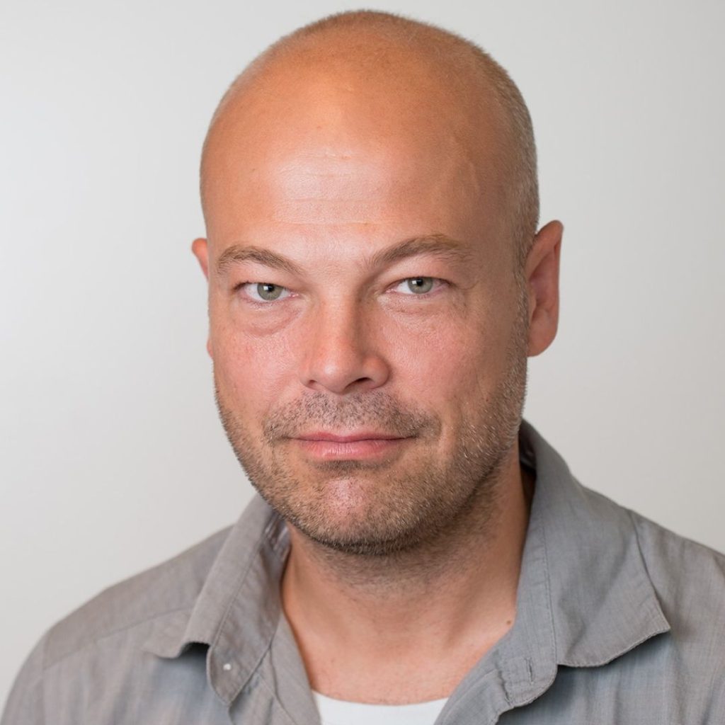
Protein stability and misfolding in keratin disorders
Rasmus Hartmann-Petersen’s project aims to characterize all possible missense variants (changes in genes which introduce a different amino acid in the resulting protein) in human keratins and investigate the importance of these variants in associated diseases.
Keratins are intermediate filament proteins that form a cytoskeletal network within cells. They are expressed in a tissue-specific fashion and form heterodimers, which then further oligomerize into filaments. Variants in several keratin encoding genes are linked to a range of hereditary disorders, including several epidermal skin diseases. On the molecular level, some pathogenic keratin variants appear to cause aggregation of the keratins.
In Rasmus Hartmann-Petersen’s project it is hypothesized that most keratin-disorders are protein misfolding diseases, i.e. diseases where the underlying genetic variants cause misfolding of the encoding protein. Rasmus and his team aim to explore this hypothesis by using computational tools, including large language models (a specific form of AI). They will test the validity of the computational predictions through focused cellular studies on selected keratins and identify components regulating keratin turnover.
The results will highlight the underlying molecular mechanisms for keratin-linked human disorders and provide predictions on the severity of all possible (both known and yet unobserved) coding variants in human keratin genes. The results could be of diagnostic value, but may also highlight the cellular protein folding and protein quality control machinery as potential therapeutic targets.
Rosaria Gandini
Assistant Professor, Aarhus University, Denmark, DKK 3.5m
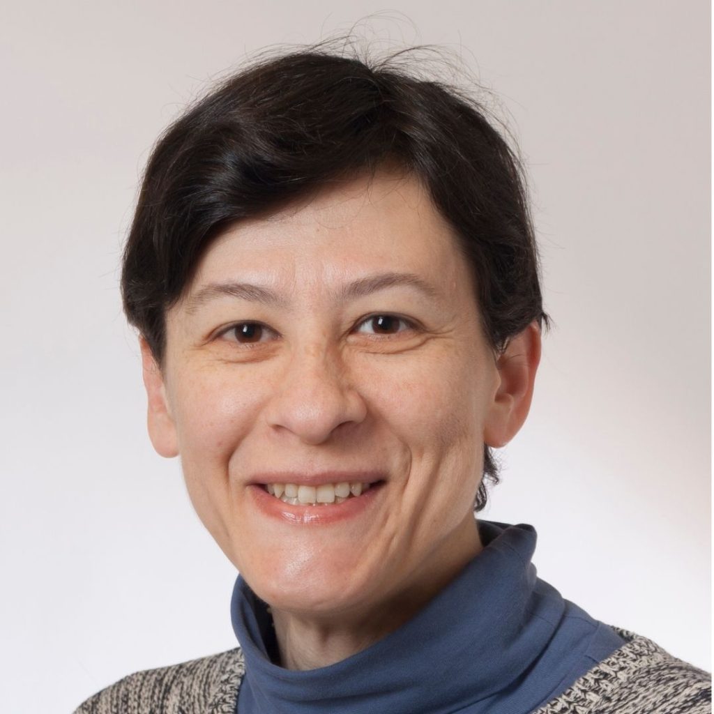
Structural dissection and dynamic insights into the molecular switch of mast cells and basophils: a blueprint for novel urticaria therapies
Rosaria Gandini’s project investigates the molecular details of the IgE-FceRI complex and its functioning on mast cells and basophils in order to improve treatment opportunities for urticaria.
Urticaria, a common inflammatory skin disorder characterized by itchy wheals, angioedema, or both, manifests in acute (AU) and chronic (CU) forms. It significantly impairs patients’ quality of life, causing sleep disturbances due to pruritus, fatigue, and anxiety. The symptoms arise from the activation of skin mast cells and basophils, leading to the release of histamine and other inflammatory mediators. This activation is initiated by cross-linking and clustering of the complexes between immunoglobulin E (IgE) and its high-affinity receptor, FceRI, which is expressed on the surface of these cells.
The FceRI-IgE complex hence acts as a powerful molecular switch, which initiates the inflammatory cascade and thus provides an attractive target for drug intervention. The structural basis of this activity, however, remains open.
Rosaria Gandini’s project aims to determine the structure of the FceRI-IgE membrane complex using state of the art Cryo Electron Microscopy (cryo-EM).
Successful elucidation of the molecular details of the entire complex and its conformations will allow identification of specific regions on FceRI for targeted intervention. This knowledge will deepen the understanding of the interaction of antibodies with Fc receptors in general and may pave the way for the development of specific and effective treatment of urticaria and related disorders.
Application and evaluation process
The LEO Foundation has established a formal evaluation process with a panel of national and international external experts from the skin research community to assist the Board in ensuring that grants are given to the best projects and the most qualified applicants.
All applications are evaluated by the LEO Foundation’s independent Scientific Evaluation Committee. Based on the evaluations by the Scientific Evaluation Committee, the final decisions on grants are made by the Board.
Next application round in 2024
The LEO Foundation calls for applications for research projects focusing on the skin and its diseases on an ongoing basis. The next call opens 1 August 2024 and closes on 12 September 2024.
The competition for Research Grants is open to talented skin researchers at PhD level or above from any country. The typical grant amount applied for is DKK 2–4 million for a period of 1–3 years. Researchers who would like to apply for a LEO Foundation research grant will be able to apply here.
Get an overview of current funding opportunities from the LEO Foundation here.
